BIOE 301D: Hands-On Microfluidics Device Laboratory
Microfluidic devices developed for biomedical research in academic labs often fall short of their intended biological and medical potential due to a persistent technology transfer gap between the engineers who design them and the biologists who are the intended users. To address this, we have developed an inquiry-based approach that leverages graduate student talent and creativity to meet the unmet needs of bioscience laboratories. Unlike many traditional fabrication laboratories in which devices created by students are discarded at the end of the course, the approach directs student efforts towards designing and fabricating devices needed by bioscience labs across campus. In the process, students experience first-hand the unexpected challenges inherent to cutting-edge research and learn to communicate across disciplines.
Winter 2025
Teaching Team: Polly Fordyce, Hawa Thiam, Jennifer Ortiz Cárdenas, Shawn Cai, Allen Yesin, Jeongwoong Yoon
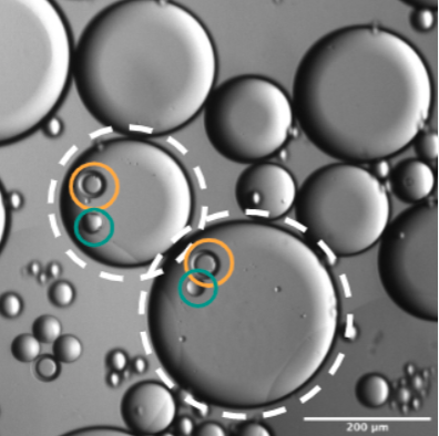
Project #1: Microfluidics trapping of single Chlamydomonas Reinhardtii cells for microscopy applications
Student team: Rong Chi, Megan Fleeharty, Kyle McLelland, Valerie Perez-Medina
Collaborators: Liat Adler (Matthias Garten Lab)
Microfluidic single-cell trapping of the green algae Chlamydomonas reinhardtii poses design challenges, as the cells are small (less than 10µm across) and deformable. Trapping them thus requires very small traps and precise flow-rates. We were able to successfully trap single cells by creating 1.5µm tulip shaped traps and flowing cells into the device at 8µL/min. We found that smaller traps were difficult to fabricate, larger traps did not result in trapping, slower flow rates resulted in cells getting stuck in the tubing, and faster flow rates resulted in broken devices and cells being forced through the traps. We were not only able to trap single cells in our devices, but to visualize organelle localization within these cells.

coming soon
Project #2: Building a Double-layer Brain Vasculature On-a-chip System
Student team: Emily Hagens, Milo Eirew, Qi Jiang
Collaborators: Sophia Shi (Carolyn Bertozzi’s lab)
The glycocalyx layer lining the lumen of endothelial cell vessels in the blood-brain barrier (BBB) has recently become an important focus of research related to aging and the study of neurodegenerative disease (Shi et al., 2025). In order to better study the development and maintenance of this layer in relation to the BBB within a lab context, we have fabricated microfluidic devices designed to model human vasculature within the BBB. Here, we show that we were successful in fabricating single-layer devices with a vessel-like structure, in which endothelial cells were able to survive for up to 10 days and form a cell layer. Additionally, we were able to construct double-layer devices in which endothelial cells and astrocytes were successfully grown in layers alongside one another, separated by a thin membrane which allowed for the exchange of small molecules between the cell types.

coming soon

coming soon

coming soon

coming soon
Project #3: Designing Microfluidic Devices for Multiplexed Single-Molecule Force Measurements
Student team: Owen Dunkley, Claudia Phillips, Sebastián Somolinos, Lauren Takiguchi, Tomasz Zaluska
Collaborators: Matt DeJong (Polly Fordyce’s lab)
Measuring biological forces at the single-molecule level is essential for understanding the out-of-equilibrium molecular dynamics that dictate the rates of life at all scales, yet current force-sensing techniques, such as optical tweezers and atomic force microscopy, lack deployability and throughput. These limitations hinder our ability to capture the complexity of biomolecular interactions in the large sequence spaces that determine their affinities. Herein, we describe the successful fabrication and experimental use of a valve-less, single-layer microfluidic polydimethylsiloxane (PDMS)-glass interface device that enables multiplexed single-molecule force spectroscopy on a standard inverted epifluorescent microscope. We undertake two design-build-test iterations on 30-chanel candidate single-layer devices built to push or suction buffer containing fluorescent ligand-bound beads uniformly between barcoded channels. Our final design addresses key issues encountered during initial device testing, incorporating taller channels and simple features to ease bubble evacuation, increasing matrix stiffness to prevent feature collapse, and optimizing channel and hole punch feature design to accommodate user variability in device manufacturing, loading, and testing.

coming soon
Spring 2024
Teaching Team: Hawa Thiam, Jennifer Ortiz Cárdenas, Christopher Choi, Heena Saqib
Project #1: Engineering Synthetic Endosymbiosis: Microfluidic Fusion of Protoplasts with Cyanobacteria
Student team: Aurora Xu, Lorenzo Magni, Sylvie Wilson, Chan-yu Kuo, Antonio Salcido-Alcántar
Collaborators: Solène Moulin (Ellen Yeh’s lab)
In some diatom and algal species, endosymbiotic cyanobacteria naturally allow intracellular nitrogen fixation, a highly desirable trait for crops. To generate synthetic nitrogen-fixing endosymbionts in plants, a system to deliver large bacteria and to screen and characterize the fusion products would be required. With its high throughput and single-cell resolution, microfluidics has the potential to enable such a platform. Here, we explored the optimization of a previously developed microfluidic-based device to trap, pair and fuse protoplast cells of two different types, with the goal of expanding this tool to protoplasts with cyanobacteria-containing vesicles. Different trap sizes, array spacing and flow rates were explored to maximize trapping efficiency.

Schematic of trapping device, middle region containing cup-trap array. Multi-inlet and outlet allow for sequential introduction of cell types and rinsing steps for efficient pairing.
Project #2: Using microfluidics to track biomolecular condensate formation
Student team: Bianca Edozie, Bianca Linden, Daniel Ilyn, Vishnu Shankar, Anjini Chandra
Collaborators: Aoon Rizvi (Serena Sanulli’s lab)
Biomolecular condensates are membraneless compartments associated with liquid-liquid phase separation, where a homogeneous solution can separate spontaneously into two distinct phases. By undergoing phase separation, cells can segregate specific proteins and nucleic acids in condensates and precisely control reaction kinetics both spatially and temporally. Biomolecular condensates have garnered attention as a pivotal player in physiological processes in eukaryotic cells, particularly in gene regulation and cell stress responses. In recent years, it has been found that these condensates form with heterochromatin protein (HP1), chromatin, and RNA by phase separation. HP1 is a highly conserved protein that is able to bind to both chromatin and RNA. Based on in-vitro experiments, the binding of HP1 to heterochromatin enables specific conformational changes to the nucleosome, exposing particular regions that can regulate gene transcription. The exact phenomenon behind the formation and dynamic behavior of these biomolecular condensates are not well understood. Within minutes of mixing equal parts of HP1-chromatin and RNA, biomolecular condensates may form. One means of encapsulating the condensates is oil-in-water droplets, where the differences in hydrophobicity results in the formation of droplets that minimize oil and water contact area and can encapsulate reagents of interest. Thus, creating a device that is able to both mix these two components to form droplets and capture the resulting droplets for imaging would help understand the mechanisms behind condensate formation, along with reaction kinetics. Therefore, we developed a two-component microfluidic device, which included the following: 1) a droplet generator to mix and encapsulate HP1-chromatin and RNA 2) a droplet trapper to trap the generated droplets and enable us to image and observe condensate formation.

Mold of droplet generator.

An L2 C. elegans worm inside the microfluidic trap.

Frontside traps with 50-50 um spacing, with ~38% trapping efficiency of two different sized protoplasts.

Succesfully trapped polystyrene beads in serpentine-style droplet trap.
Project #3: Immobilization of C.elegans for tracking sex myoblast cell migration using a microfluidic device
Student team: Rucha Bhise, Kimberly Cheung, Ezra Haddad, Allen Yesin, Jeongwoong Yoon
Collaborators: Ayaka Kasamatsu(Jessica Feldman’s lab)
Immobilizing Caenorhabditis elegans (C. elegans) larvae for long-term imaging is crucial for studying cellular migration, particularly the migration of sex myoblast (SM) cells. This study focuses on developing a microfluidic device to immobilize L2 stage C. elegans for tracking SM cell migration towards the center of the gonad over a four-hour period. The device, designed to fit on a standard cover slip, incorporates microchannels that taper to match the worm's dimensions, preventing rotation and ensuring proper orientation during imaging. Multiple design iterations were tested, focusing on channel dimensions, flow rates, and feeding mechanisms to optimize worm immobilization without hindering feeding. The final design, with 10 µm height channels tapering from 20 µm to 7 µm, successfully immobilized L2 worms while allowing normal SM cell migration, observed through fluorescence imaging.

Fluorescently labelled sex myoblasts migration was tracked over ~40 minutes.
Winter 2022-2023
Teaching Team: Polly Fordyce, Hawa Thiam, Jennifer Ortiz Cárdenas, Hope Leng, Luis Santiago Mille, Daria Wonderlick
Project #1: Microfluidic generation of single and double emulsion droplets to encapsulate large cells
Student team: Andy Hung, Annie Nguyen, Tianyu Lu, Maya Sheth
Collaborators:
While droplet microfluidics has proven to be a powerful tool for single-cell analysis, the commercially available 10X Genomics droplet generator has limitations in accommodating large cells, as their maximum supported cell size is 30 μm in diameter. The maximum theoretical cell size as determined by the cell channel dimension is 60 μm. To address the need of single-cell sequencing and other droplet-related assays for cells beyond 60 μm in diameter, we designed and tested proof-of-concepts for microfluidic single and double emulsion droplet generators that can accommodate cells or other objects up to 250 μm in diameter. We developed a single emulsion droplet generation system that generated droplets in the range of 240 to 500 μm in diameter aimed to encapsulate both large cells and barcoded beads, making it a valuable tool for single-cell analysis. We also developed a double emulsion droplet generation system that employs a two-step process to encapsulate large cells. By controlling the flow rates of the three phases involved in the process, we were able to generate uniform double emulsion droplets with a mean inner diameter of 240 μm and mean outer diameter of 360 μm.
Co-encapsulation of double emulsion droplets (orange) and MRBLEs beads (blue) in single emulsions.
Project #2: A Microfluidic Device for Long-Term Imaging of Gut Repair in Mutant L1 C.elegans larvae
Student team: Christopher Choi, Emily Gardner, Kenna McRae, Carolina Rios-Martinez, and Heena Saqib
Collaborators: Lauren Cote (Jessica Feldman’s lab)
Caenorhabditis elegans (C. elegans) are free-living transparent nematodes that live in temperate soil environments. Recently, Jessica Feldman’s lab at Stanford along with the Pickett lab at SJSU have discovered an exciting model for congenital repair of damaged epithelial tubes using the C. elegans intestine. The conserved scaffold protein PAR-3 is responsible for the polarization of epithelial cells into distinct apical, lateral, and basal cortices, which is essential for epithelia to act as selective barriers. Preliminary data has shown that PAR-3 depleted C. elegans worms die before hatching from eggshells due to disconnected epithelial cell walls. Nevertheless, worms with gut specific apical PAR-3 depletion were able to repair their cystic guts through re-expression of PAR-3. We developed microfluidic devices that can trap and align 40 mutant L1 C. elegans larvae at once for long term imaging, restricting the activity of the mutant worms without the need to introduce immobilizing drugs. The trapping mechanism was optimized by implementing and testing the dimensions of a wide entrance region (fixed at 20µm), a narrow region, a trapping region, and the final taper region. We successfully performed long term imaging (72 hours) in which no worms escaped the traps and remained alive and aligned. We were also able to observe distinct repair of mutant L1 C. elegans worms at the end of the session.

Schematic of trapping mechanism.

Double emulsions trapped in U-trap device.

Flow and shear stress simulations of single and dual biofilm growth chambers.
Encapsulated plant protoplasts engineered to constitutively express GFP from large Nicotiana benthamiana leaves in single emulsion droplets.
C. elegans at end of 10 µm trapping region.
Project #3: Trapping double emulsion droplets for time-lapse imaging
Student team: Nicolai Dorka, Ishan Gaur, Douglas Henze, Jessica Karaguesian, Ernst Pulido
Collaborators: Ali Lashkaripour (Polly Fordyce’s lab)
Trapping individual single and double emulsions over time bears great potential for high-throughput screening with time-resolved information. Nonetheless, most droplet-based assays rely on end-point measurements either through on-chip sorting or off-chip FACS sorting (for double emulsion droplets). Therefore, information regarding dynamics and kinetics is often lost in droplet-based assays. To this end, the ability to image individual droplets over time enables the observation of transient responses. We developed two chip designs that enable time-resolved high-throughput screening of double emulsions: microwells and U-traps. Trapping double emulsions with microwells had several advantages over U-traps. First, microwells had a higher trapping efficiency, making them more suitable for valuable and less abundant samples. Droplet trapping in U-traps rely on collision to break the snake flowing pattern around the traps. This requires a higher droplet density compared to the microwell design. Second, double emulsion droplets remain captured in microwells with or without flow, whereas U-traps need a constant flow rate, which makes microwells the preferred choice for transport and set up changes (e.g. switching between imaging modalities).

Double emulsions trapped in microwells device.
Project #4: Long-term Biofilm Imaging Using a Microfluidic Device
Student team: Hajime Fujita, Xiangmeng Shawn Cai, Isabel Goldaracena, Emilie Kono, Deepak Gopalan
Collaborators: Soumaya Zlitni (Ami Bhatt’s lab)
Biofilms are assemblies of microbes on a surface, embedded in an extracellular matrix that act as a protective barrier for the microbes, shielding them from the external environment. This means that bacterial biofilms are much more resistant to antibacterial treatments than planktonic bacteria and hence a common cause of persistent infections, putting a heavy burden on our healthcare system. However, most currently used assays are unable to study and quantify biofilm formation under an environment that is close to the natural physiological conditions. We developed a microfluidic device that allowed for long-term, un-disturbed bacterial biofilm formation. We fabricated different designs having different growing chambers and have the flexibility of having more replicates per experiment. Using these devices, we were able to grow bacteria (E. coli and E. faecalis) in a controlled environment (constant flow, temperature, nutrients) and observed bacterial growth and biofilm formation in the device over time.

Fluorescence signal immediately after the bacteria were injected (top, t = 0 hrs) and after overnight incubation (bottom, t = 24 hrs).
Winter 2021-2022
Teaching Team: Polly Fordyce, Jennifer Ortiz Cárdenas, Eliel Akinbami, Renee Hastings, Ali Lashkaripour
Project #1: Droplet Encapsulation of Oil-producing Algae
Student team: Kyle Denton, Mengdi He, Hope Leng, Cyrus K., Daria Wonderlick
Collaborators: Ellen Yeh (Yeh Lab)
The microalgae Botryococcus braunii is a promising source of biofuels since it produces hydrocarbon oils that are readily converted into petroleum-equivalent fuels and the algae stores them in the extracellular space where they are more readily extracted, addressing two energy-intensive and costly steps in biofuels production. Interest in B. Braunii as a source of alternative biofuels creates the need for the development of screening processes that can identify highly efficient mutants. Our team used microfluidic droplet generators to encapsulate single algae colonies for observation of growth and quantification of hydrocarbon production. We designed and fabricated single- (SE) and double-emulsion (DE) droplet generators to encapsulate single colonies. The 50 µm inner/70 outer µm DE (water-oil-water) droplets were sufficiently big to encapsulate entire algae colonies and remained stable for 3-weeks. We also fabricated visualization chambers to enabled long-term tracking of DE droplets. These devices required little technical expertise to operate, and are suitable for any lab interested in elucidating new insights into cyanobacterial biology, e.g. metabolic pathways, photosynthetic efficiency.

Encapsulating single B. braunii in water/oil droplets.

Growing B. braunii colonies within water/oil/water droplets.
Project #2: Microfluidic Platform for Size-Based Killifish Oocyte Sorting
Student team: Ziad Ali, Ananya Chadha, Vincent Cornelius, Danielle Klinger, and Cassandra Villicana
Collaborators: Jingxun Chen (Brunet lab)
Reproductive aging can be studied by exploring the changes in oocyte (unmatured eggs) quality. The African turquoise killifish is one of the shortest-lived vertebrates used in laboratory studies, with a lifespan of ~ 6 months. Killifish exhibit signs of aging and age-related diseases during this short lifespan making them a powerful model organism to study reproductive aging. Oocyte size can vary by 2 orders of magnitude, depending on the age, with cells as small as 10 µm and as big as 1 mm. While methods like 10X Genomics enable capture and analysis of the ~ 10 - 50 µm cells, the 50 - 200 µm cells are missed by this method and thus understudied. To address this challenge, we designed and fabricated a pinched cross-flow microfluidic device to filter cells outside of the target size range. A flow ratio of 1:4 for the sample:buffer was found to be most successful in producing flow lines that divert the small cells toward the filters, and a filtration gap of 30 μm demonstrated the highest sorting ability without cell lysis. The device improved the enrichment of cells within the target size range by approximately 2.5x with each run. This suggests that through repeated filtering of cells, these devices may be useful in extracting these Killfish oocytes cells allowing examination of their genetic profiles.


Composite image over 100 minutes showing stuck “sticker” caulobacter within the first chamber.
Composite image over 1.5 seconds showing highly motile “swimmer” caulobacter within the second chamber.

Project #3: Sorting Muscle Stem Cells from a Heterogeneous Cell Suspension Using Cross-Flow Filtration
Student team: Kevin Chen, John Klich, Soham Sinha, Jonathan Weiss, Jerry Yan
Collaborators: John Eugenis (Rando lab)
Despite advances in single cell sorting, isolating specific cell populations from heterogeneous samples of digested tissue remains a prohibitively expensive and time intensive process. Sorting cell suspensions by cell type is required for both fundamental research and therapeutic translation, including isolation of muscle stem cells for regenerative medicine. We developed a cross-flow filtration microfluidic device capable of separating particles in different size regimes (<10 μm vs. >10 μm) in a bead-based model system. In an iterative approach, we demonstrated that sorting efficiency is a function of interpillar spacing and pillar angle; the most successful design consisted of 10 μm spacing between pillars, 60 degrees pillar angle, a reduced flare, and operated at a flow rate of 10 μL/min. Using this device, we achieved separation of 75% of the target 6 μm beads through the side channels and nearly 100% retention of the large beads in the main channel.

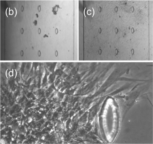
Project #4: Microfluidic Cytometer to Separate and Image Stalked and Swarmer Phase Caulobacter
Student team: Netra Unni Rajesh, Joy Doong, Linda Yang, Zi Yi Stephanie Huang, Luis Santiago Mille
Caulobacter crescentus, a freshwater bacterium, is used as a model organism to study asymmetric cell division dynamics in response to environmental fluctuations. These organisms have two distinct phenotypes: a stalked phenotype that adheres to surfaces and a swarmer phenotype with high motility. The separation of these phenotypes requires multiple rounds of density gradient centrifugation in a cold room, rendering it a complex process. We designed a microfluidic device that contained two chambers with independent media inlets, in which the separated stalked and swarmer cells could be cultured. In addition, the stalked culture chamber contained a gradient generator to enable the study of environmental fluctuation on this phenotype. We optimized the channel dimensions, 200 µm width and xxx µm length, in the gradient generator to ensure adequate mixing. We also developed a protocol that allowed for adhesion of the stalked cells, followed by a rinse that allowed the swarmer cells to be flown into their corresponding chamber. Once the phenotypes were separated, the swarmer cells were clearly adhered to the surface and cell division was observed over a 100-min imaging event. In the swarmer chamber, the cells appeared to be highly mobile. Future work will focus on deciphering if changes in C. crescentus cell cycle and a range of morphologies can be achieved by applying gradients in concentrations of antibiotics, sugar, and/or nutrients.


Round 1: Main channel of cross-flow filtration device showing a large target cell remaining in the channel while a smaller cell gets pushed through pillars and lysed.
Round 2: Target cells persisted in the main channel of the cross-flow filtration device without lysis after the between-pillar distance was reduced to 30 µm.
Image of fabricated wafer with 15 µm separations between pillars.
Image of microscope with fabricated device and tubing.
Photo showing device with generated gradient.
Winter 2019-2020
Teaching Team: Polly Fordyce, Nicole DelRosso, Louai Labanieh, Zachary Sexton
Project #1: Placenta-on-chip
Student team: Anthony Agbay, Ashwin Ramachandran, Carly Weber-Levine, Mustafa Fattah, Vandon Duong
The placenta plays an integral role in the exchange of endogenous and exogenous materials between a mother and the fetus. During pregnancy, blood from the mother passes through the placenta, where oxygen and other nutrients are filtered into the fetus’ blood supply via the umbilical cord. In the placenta, the mother and fetal blood do not actually mix, but rather come into very close contact across a thin membrane, known as the placental membrane. We sought to mimic the architecture and filtering function of the placenta by designing a placenta-on-a-chip (POC) composed of two distinct compartments, maternal and fetal, separated by a polycarbonate membrane. Using soft lithography, we were able to develop a device that can be used as a scaffold to mimic the filtering function of a placenta. The two layered device was separated by a polycarbonate membrane sandwiched between two laser cut glass slides and allowed to take in two separate inputs and release two separate outputs. We were able to visualize the HUVEC cells flowing into and through the channel, settling on the maternal side of the membrane only, and that the media flowed on the fetal side with no cross-contamination/transfer of cells from the maternal side. In future iterations of the POC we hope to obtain a fully functioning microfluidic filtration device that can additionally separate plasma from cells in blood.

Schematic showing 4 individual components that create the two-layered placenta-on-chip

Assembled placenta-on-chip device with an integrated polycarbonate membrane
Project #2: Immobilization of L2 C. elegans for live cell imaging (collaboration with James Ferguson from Jessica Feldman’s Lab )
Student team: Nora Enright, Jesse Gibson, Akshay Maheshwari, Emily Meany, Cameron Park
C. elegans is a small transparent nematode commonly used as a model organism. Its transparency throughout its lifespan enables live confocal imaging, offering important insights into cell migration and development. Previous microfluidic devices have successfully trapped and immobilized adult or stage L3-L4 worms for imaging purposes, but none have been used with stage L1-L2 worms. We have developed a microfluidic device to immobilize C. elegans in these earlier larval stages, optimizing our tapered channels to accommodate their small size. We successfully loaded worms in many of our devices using a syringe pump and determined the 15 μm tall, 20μm inlet and 5μm outlet device to be best for immobilizing the target worm population. Confocal microscopy imaging revealed successful worm immobilization over 8 hours, facilitating tracking of sex myoblast cell migration patterns in stage L2 C. elegans.

Schematic of ’clamp’ device. Parallel tapered channels with a gradual decrease in width over their length are designed to trap the worms.

An immobilized L2 worm (a) without any applied flow rate and (b) with a 1 μL/min flow over eight hours, the fluorescently labeled sex myoblast cells (green) were tracked over the course of eight hours
Project #3: Measuring red blood cell’s deformability with constriction channels (collaboration with Elizabeth Egan’s lab)
Student team: Eliel Akinbami, Jason Herrman, Eerik Kaseniit, Manish Ayushman, Sauradeep Sinha
Malaria is a life-threatening disease caused by parasites that are transmitted to people through the bites of infected female Anopheles mosquitoes. When a person is bit, the malaria parasites must get inside the dynamic membranes of red blood cells (RBC) in order to begin the infection and it has been shown that variations in the physical properties of RBC’s affect the rate of parasite infiltration. Thus, we set out to design and test a microfluidic device that could characterize the “squishiness” or deformability of RBC’s and compare their susceptibility to infection by using samples derived from patients with different genotypes. We designed two types of constricted channels, based on existing literature. As RBCs flow through the constrictions, they were deformed and the deformability of the cells determined which paths the cells take downstream. One design utilized an array of wedge constrictions and channels of various dimensions. The second design had a branching tube configuration. One major challenge of this fabrication was carefully calibrating the light doses required to achieve feature resolution of nearly one micron. A second challenge was the need for two-level fabrication to form bypass channels that were much larger than the constricted channels. In order to accomplish this, we used a multi-mask process, in which one mask defines the constricted channels, and another mask, aligned to the first using specialized markings, defines the large bypass channels. We were able to push the limits of feature resolution on these chips with small feature sizes on multiple layers. We measured our devices using a flow control system, by pulsing the RBC flow through the microfluidic device, and ultimately succeeded in observing RBCs transiting the constrictions.

Project #4: Single layer microfluidic devices to trap single mammalian cells for long-term culture and imaging (collaboration with Taihei Fujimori from Lacra Bintu’s lab)
Student team: Peter Suzuki, Taylor Merkel, Gaeun Kim, Namrata Anand, Taylor Nguyen, Callan Monette
Chromatin regulation plays an essential role in development, aging, and disease. Previous work in the Bintu lab has highlighted the importance of studying chromatin regulatory dynamics at single-cell resolution, which can be done through live-cell imaging: single-cell imaging can help characterize temporal dynamics as well as cell-to-cell heterogeneity. While live single-cell imaging is common in adherent cells, we are still quite limited in our ability to perform single-cell tracking of suspension cells over a long period of time. Furthermore, trapping of suspension cells presents the opportunity to release cells from their traps for collection and downstream analysis. We aimed to design a microfluidic device capable of trapping and immobilizing hundreds of single cells such that they can be maintained over the course of a 5-day live cell imaging experiment. We conceptualized two different single-layer design approaches: a plinko-style lateral trapping device, and a serpentine capture device. We successfully achieved a high capture efficiency for both devices, and incorporated two additional modular devices capable of (1) filtering debris that may be introduced during media flow and (2) enabling switching between cell suspension and media flow with minimal introduction of bubbles. In the future, parameters such as gap width and length could be optimized to avoid cell escape over time. Further changes, such as using multiple syringe pumps in a given experiment rather than swapping a syringe mid-experiment, are needed to enable longer-term culture without introduction of bubbles or dislodging of cells.

Schematic of serpentine and plinko-style trapping devices.
Winter 2018-2019
Teaching Team: Polly Fordyce, Loza Tadesse, Peipei Lyu
Project #1: Shear-free oligodendrocyte culture with media exchange (collaboration with Manasi Iyer from Brad Zuchero’s lab)
Student Team: Beatriz Atsavapranee, Patrick Brennock, Nicole DelRosso, Eva Gonzalez Diaz, & Connor Ludwig
Myelination is essential for proper brain function, and dysfunction in the process is linked to several neurological disorders. Oligodendrocytes are glial cells that myelinate neuronal axons in the local environment and contribute to brain plasticity. These cells and their myelination dynamics are difficult to study due to their shear sensitivity, precise imaging requirements, and limited throughput. To address this, we designed, fabricated, and tested microfluidic devices to culture oligodendrocyte progenitor cells, differentiate oligodendrocytes, and image signaling dynamics on-chip with minimal shear stress. We successfully fabricated devices compatible with high-throughput confocal imaging, and seeded and cultured cells on-chip for up to 7 days under perfusion. In addition, we determined the maximum flow rate to reduce background calcium transient signals, and demonstrated the ability to rapidly exchange fluids on the order of seconds. These results show that microfluidic devices are a promising platform to study the impact of neuronal activity on myelination with a streamlined, on-chip workflow that minimizes shear stress.
“Horseshoe device” cell loading: day 1

“Horseshoe device” cell loading: day 7
Loading cells into ‘horseshoe devices’.
Project #2: A hydrodynamic breadboard (with Endre Mossige and Chunzi Liu in Gerry Fuller’s lab)
Student Team: Michaela Hinks, Prima Dewi Sinawang, Zachary Sexton, Ana Uriarte, & Sasha Zemsky
Sorting cells represents a critical need in cellular and molecular biotechnology, medicine, and tissue engineering. Although technologies such as fluorescence activated cell sorting (FACS), magnetophoresis, and optical tweezers have been widely implemented for cell sorting, these methods also require cell labeling, extensive hardware, and may only be efficient at sorting cells for short periods or in small batches. Furthermore, when sorting cells based on size selection, these methods may not be the most effective approach. Traditionally, sorting cells based on size has been a passive process involving filtration, physical force, or tailored surface chemistry methods to direct cell separation. While microfluidic devices have leveraged these passive physical properties to overcome the gap in sorting accessibility, these devices lack modularity and universal applicability without extensive design modification. Here, we present a device capable of creating a hydrodynamic pillar in which the diameter of the pillar can be adjusted by simply varying flow rates of the source and sink of the microfluidic device. Furthermore, no clogging is observed along the boundary of this hydrodynamic dipole pillar, making it possible to use deterministic lateral displacement to sort particles and cells with a wide working range. Our results show that this preliminary device is able to sort particles with diameters of 5 μm and 10 μm.

Pneumatic and imaging setup for hydrodynamic breadboard.
Hydrodynamic barrier (red) in the midst of cross-flow (blue).
Differential flow of fluorescent particles around the hydrodynamic barrier.
Project #3: High-throughput production of artificial cells (with Akshay Maheshwari in Drew Endy’s lab)
Student Team: Amy Bour, Suzanne Calhoun, John Eugenis, Yuxi Ke, Alice Stanton
Liposomes are aqueous vesicles surrounded by a lipid bilayer that are broadly used in basic science research, drug delivery, and other applications. Liposomes are typically created in bulk by mixing aqueous and oil solutions, but this approach is time- and reagent-intensive and produces polydisperse emulsions. Here, we harness microfluidics to create a droplet generator that can produce liposomes with more controllable properties and using smaller volumes of reagents. Hydrodynamic flow focusing directs multiple streams to create concentric liposome layers in a microfluidic device. We report the creation of small, cell-sized (2.5-30um) unilamellar liposomes for synthetic biology applications using a device that can be used to form double emulsions enclosing various cell components. This can enable future work creating artificial cells and further our understanding of the role of various cellular components comprising living cells. The tunability of liposome properties, small achievable liposome sizes, and small reagent volumes make this device broadly attractive for a wide variety of uses in basic science research and other delivery applications.

Image of small (5 µm) features at device nozzle.

Polydisperse double emulsion droplets with sizes ranging from 2 µm to 30 µm.
Production of small single emulsion droplets.
Project #4: A simplified device for collagen fiber production
Student Team: Ethan Li, Abhijit Lavania, Jennifer Moy, Elizabeth Botbol Ponte, Grace Zhong
Collagen is an indispensable structural protein in vertebrate extracellular matrix and is the material of choice for many biomedical applications due to its superior physical properties and biocompatibility. In particular, aligned collagen is of great interest in engineering scaffolds for tissue regeneration. However, current fiber extrusion methods are not tunable, have a large lower diameter limit, and produce fibers with inferior mechanical properties. Here, we describe the design and fabrication of various single- and multi-layer devices for collagen extrusion. We explore the effect on fiber formation from factors such as priming sequence, junction geometry, channel length, single-vs. multi-layer designs, and flow rates. We further present an optimized priming sequence which enables successful fiber extrusion.

Bright field image of 2 layer flow-focusing device.

Bright field image showing co-axial flows.
Successful formation of collagen fibers at device outlets!
Collagen fiber formation within device channels.
Winter 2017-2018
Teaching Team: Polly Fordyce, Siavash Ahrar, Yuan (soso) Xue, Alec Tarashansky
Project #1: A planarian guillotine
Student Team: Bauer LeSavage, Jack Silberstein, Jon Calles, and Suhas Rao
The planarian flatworm S. mediterranea is a unique model of development that can regenerate entire organisms from fragments as small as 1/279th of the original host. This extreme regenerative capacity is thought to result from the planarian’s high stem cell content (up to 30% of total cells). Chimeric worms created by fusing fragments from different worm species provide a powerful tool to study the collective action and coordination of these stem cells in regenerating an entire organism. This competition is best studied by bisecting worms along their longitudinal axis and fusing halves from opposite strains together; however, this requires the ability to consistently and efficiently bisect worms with a throughput and precision not possible via current methods. To address this, we developed a microfluidic guillotine to quickly and consistently bisect planaria and recover the halves for further regeneration studies. Our device consists of an 18 mm long, 175 µm tall, and 700-900 µm wide channel ending in a sharp PDMS blade (6 degree angle) such that worms are flowed down the channel, collide with the blade and are bisected into two pieces, with each half flowing out of its own outlet channel. We demonstrated that individual worms could be bisected and that bisected worms were viable and capable of future regeneration.

Figure 1. Schematic showing microfluidic planarian guillotines with varying channel widths after the guillotine blade.

Figure 1. Device design schematic.
Video 1. High speed video showing bisection of a single planarian using the microfluidic guillotine.
Project #2: a device for long-term culture of C. elegans
Student Team: Prashanth Srinivasan, Pengyang Li, Spencer Cesar
The free-living nematode Caenorhabditis elegans is a model organism for the study of changes during aging. Recently, several studies have reported that pheromones secreted by male worms cause reduced lifespan and accelerated aging in hermaphrodite (her ) worms. However, the identity of these pheromones and their mechanisms of lifespan reduction are unknown. Current methods for studying sexual interactions in C. elegans are laborious, time-intensive, and error-prone because they require manual separation of sexes and removal of progeny. Moreover, the low throughput of manual worm separation precludes the study of context-dependent pheromone secretions. A previous attempt to maintain sex separation using a microfluidic device relied on failure-prone screw valves and a single-channel loading strategy susceptible to cross-contamination of the sexes. Here we report the design, fabrication, and initial testing of a valve-free microfluidic device capable of maintaining male and her worm populations in chemical contact but physical isolation for probing the molecular mechanisms of pheromone-mediated lifespan reduction. The final device should also satisfy following criteria: able to keep 10-20 worms for 21 days (each sex); eggs laid can be removed periodically; worms in the device can be fed with E.coli ; and transparent enough to monitor and image the worms.
Figure 2. Hermaphrodite and male C. elegans worms swimming in microfluidic device.
Project #3: A valved device for co-culture of macrophage and bacteria
Student Team: Peipei Lyu, Andres Aranda-Diaz, Punnag Padhy, and Thomas Lozanoski
When a macrophage becomes infected with or phagocytoses a bacterium, (e.g. E. coli or S. typhimurium), the infected cell engages in a coordinated inflammatory program to destroy the pathogen; however, the response is not always successful, and sometimes the pathogens evade the immune system, leading to severe infections. Understanding the mechanisms that govern immunosuppression will help medical scientists develop effective vaccines and treatments for problematic infections. One hypothesis for immunosuppression is that the macrophages that phagocytose the bacterium are inhibited from relaying inflammatory signals to their neighboring immune cells. Current methods for studying bacterial infection of mammalian cells with live-cell imaging do not enable the response to infection to be separated from responses due to paracrine signaling. For example, conditioned media only captures signaling at one time point and does not lend itself to studying feedback between infected and non-infected cells. To address these issues, we microfabricated a device capable of spatially and temporally separating different cell populations so that the infection process can be imaged and the effects of its downstream signaling can be measured.
Movie 1. Pneumatic valve actuation.

Figure 1. Fluorescence image showing macrophages (green) and E. coli (red) within a chamber of the microfluidic device.
Project #4: A microfluidic droplet generator for testing polymer hydrogels
Student Team: An Ju, Loza Tadesse, Xinzi Wang
Hydrogel microspheres have been shown to enable encapsulation of cells and other biomolecules and reduce shear damage specially for injecting to an organism. Here, we investigated pre-encapsulation of mesenchymal stem cells in hydrogel microspheres using a microfluidic platform. We fabricated and tested two droplet forming designs (T-junction and flow-focusing), investigated hydrogel types including their polymerization parameter, and tested cell loading behavior of the droplets.
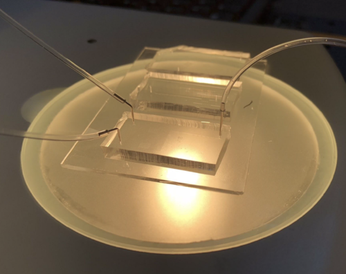
Figure 1. Microfluidic droplet generator connected to syringe pumps on microscope illumination stage.

Figure 2. Droplets produced via T-junction (top) and flow focusing (bottom) devices at different flow rates.
Winter 2016-2017
Teaching Team: Polly Fordyce, Kara Brower, & Diego Oyarzn
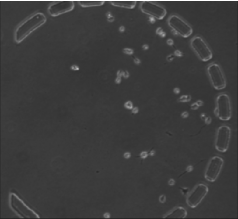
Project #1: A microfluidic device for studying toxoplasma gondii
Student Team: Sam Bray, Cooper Galvin, Ali Hemmatifar, Deze Kong, and Tim Schnabel
Extravillous trophoblasts (EVT) grow out from the fetal placenta and invade the maternal uterus. While the establishment of EVT cells in the uterus is a vital stage in establishing pregnancy, the rarity of these cells has made them difficult to study. Furthermore, some EVTs differentiate into extremely large (>100 ɥm) and polyploid (>100 N) cells and we know little to nothing about the functions of these unique cells. To address this issue, we attempted to construct a microfluidic device with physical barriers for sorting and capture of large EVTs. We have engineered a single-layer microfluidic device to capture cells using physical separators at defined widths (90, 75, and 50 um). Using reverse flow, we selectively captured objects of defined diameters from individual chambers. This device will allow for the isolation of large EVTs for subsequent downstream analysis and functional studies (RNAseq, proteomics, motility studies).
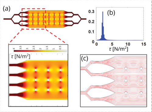
Figure 1. Shear stress modeling results from COMSOL and particle image velocimetry.
Project #2: Placentaur: a device for isolating very large cells from a cell mixture
Student Team: Alexander Tarashansky, Terence Theisen, Shreya Deshmukh, Nelson Hall, and Yuan Xue
Extravillous trophoblasts (EVT) grow out from the fetal placenta and invade the maternal uterus. While the establishment of EVT cells in the uterus is a vital stage in establishing pregnancy, the rarity of these cells has made them difficult to study. Furthermore, some EVTs differentiate into extremely large (>100 ɥm) and polyploid (>100 N) cells and we know little to nothing about the functions of these unique cells. To address this issue, we attempted to construct a microfluidic device with physical barriers for sorting and capture of large EVTs. We have engineered a single-layer microfluidic device to capture cells using physical separators at defined widths (90, 75, and 50 um). Using reverse flow, we selectively captured objects of defined diameters from individual chambers. This device will allow for the isolation of large EVTs for subsequent downstream analysis and functional studies (RNAseq, proteomics, motility studies).

FIgure 1. Device schematic. (A) Each separation chamber has different pillar spacings to capture cells of different sizes. (B) Integrated valves allow selective retrieval of particular cell sizes.
Figure 2. Example images showing overnight growth of cells.
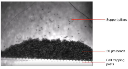
Figure 2. Image of the microfluidic device containing pillars with 50 µm gaps between them and 50 µm diameter beads successfully captured under constant pressure flow.
Project #3: Traptasia: Devices to capture and study the cnidarian apitasia
Student Team: Salil Bhate, Daniel Hunt, Louai Labanieh, Sarah Lensch, and Will van Treuren
Coral reefs are some of the most productive ecosystems on earth. Driving this productivity is a symbiosis between corals (phylum Cnidaria) and dinoflagellates (predominantly in the genus Symbiodinium) where the coral provides inorganic nutrients and the algae provides glucose or other fixed carbon. Coral bleaching, the phenomena where coral eject their symbionts when stressed, is a major concern for coral reefs around the globe. Unfortunately, coral reefs are currently threatened by systemic anthropogenic stressors, primarily ocean warming and acidification. Understanding the mechanics of coral bleaching is essential to responding to this threat. Here, we develop a microfluidic device that enables much more statistically rigorous quantification of coral biology in a model system, increasing the length of viable experiments by a factor of 3 and the number of replicates by a factor of 50.

Figure 1. Image showing array of traps with Aiptasia larvae within them.
Movie 1. Trapped Aiptasia larvae expelling symbiotic algae under stress.
This project was continued by some of the team members after the course finished, leading to a manuscript currently under review.
Please see the full bioRXiv post here!